Orbital Roof Fracture Ct

Orbital Roof Blow In Fracture Radiology Case Radiopaedia Org

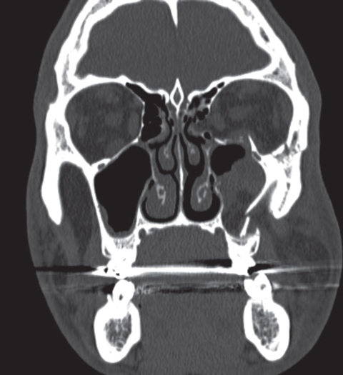
Superior Orbital Roof Blowout Fracture With Intact Orbital Rim Radiology Case Radiopaedia Org

Orbital Blowout Fracture Radiology Reference Article Radiopaedia Org

Pin On Ct Scans
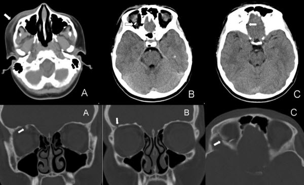
Orbital Roof Blow In Fracture A Case Report And Review Jones Journal Of Radiology Case Reports

Multidetector Ct Of The Pelvis Shows A Fracture Dislocation Of The Hip Red After Car Accident Normal Is Gre Radiology Diagnostic Imaging Radiologist

Orbital roof fracture icd 801 01 etiology.
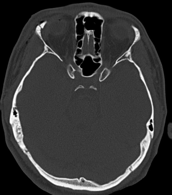
Orbital roof fracture ct.
Zygomatic Process Of Frontal Bone Fracture Radiology Case Radiopaedia Org

Pin Pa Ct Scans

Fd Surfer S Ear Acquired Bony Exostoses Of The External Auditory Canal Ear Surfer Head And Neck

Superior Orbital Fracture Radiology Case Radiopaedia Org

Fibrous Dysplasia Frontal A And Lateral B Plain Radiographs Show A Well Defined Thickened And Sclerotic Appearance Of The R Patient Radiology Radiographer

Extremely Large Ruptured Abdominal Aortic Aneurysm Aortic Aneurysm Abdominal Aortic Aneurysm Aneurysm

Capito Hamate Coalition Radiology Case Radiopaedia Org Radiology Bone And Joint Bone Diseases

Ct Findings Of Stercoral Colitis Dilatation 6 Cm And Wall Thickening 3 Mm Of The Affected Colon Segment Pericolonic F Segmentation Colitis Celestial Bodies

Medpix Case Blowout Fracture Of Orbit

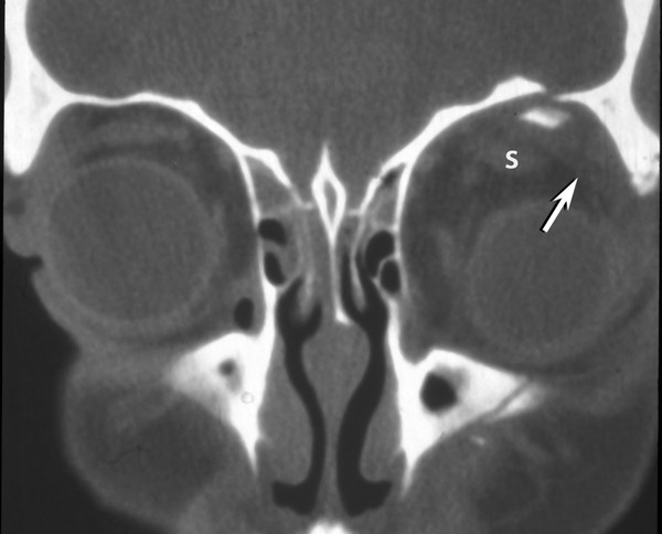
Osteoma Of Orbital Roof Radiology Case Radiopaedia Org Radiology Radiology Imaging Diagnostic Imaging

Blowout Orbital Fracture Radiology Medical Imaging Radiography

North Broward Radiologists Radiologist Radiology Mri

Radiology Rounds Radiology Rounds Medical Specialties Medical Training Radiology

Vocal Cord Paralysis Radiology Reference Article Radiopaedia Org In 2020 Thyroid Surgery Paralysis Nerve Palsy

Copper Beaten Skull Also Known As Beaten Brass Skull Refers To The Prominence Of Convolutional Markings Gyral Impressions On The Inner Table Of Th Tip Beyin
15 Year Old Male With Occular Trauma Axial Ct Shows Fracture Of The Download Scientific Diagram

Pin On Mr Radiology

Radial Head Fracture Radiology Case Radiopaedia Org Radiology Fractures Medical School Stuff
1

Pin En Mri

The Atlantodental Interval Adi As The Name Suggests Is The Horizontal Distance Between The Anterior Arch Of The Atlas And The Radiology Intervals Occipital

Facial Bone X Ray Lateral View Www Anatomynote Com Facial Bones Radiology Radiology Student

Pin By Dr Abuaiad On Brain Head And Neck Thyroid Cyst Thyroid Gland Skin Issues
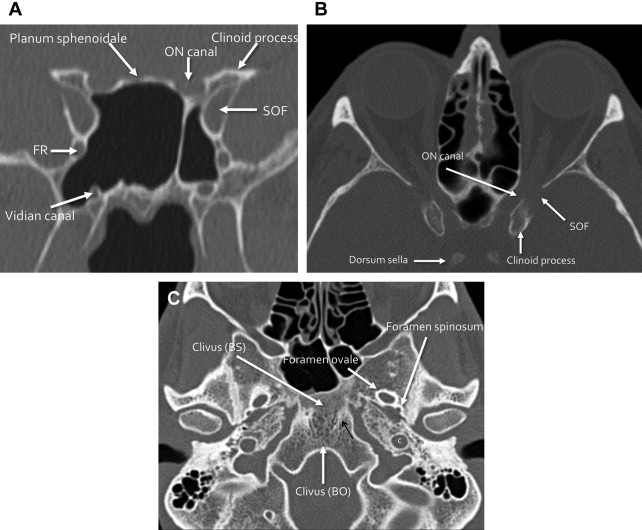
Skull Base Fractures And Their Complications Radiology Key

Burst Fracture Of Lumbar Spine Sagittal Reconstruction Of Ct Of The Lumbar Spine Demonstrates A Comminuted Vertical Burst Fracture Through The Body Of L1 Whit

The True Stories Of A Rapper Whose Lungs Collapse Photos Lunges Healthy Lungs Ct Scan

Foot Fractures In Adults Radiography Broken Foot X Ray
Traumatic Orbital And Occular Injury Radiology Key

Fd Lymphatic Intravasation This Hsg Shows A Streaky And Mesh Like Collection Of Contrast Material In The Right Side Of Th Lymphatic Material Radiology

Bulley Sign Radiology Bullseye View The Bullseye View Is Designed For Better Evaluation Of Lesion Located In Retroareolar Are Red Bone Marrow Signs Radiology

Plasmacytoma Radiology Case Radiopaedia Org Radiology Case Limb

Sacrococcygeal Giant Cell Tumour Radiology Case Radiopaedia Org Tumor Radiology Cell

Soap Bubble Lesions Caused By Cryptococcus Neoformans Radiology Nuclear Medicine Neurology

Subependymal Giant Cell Astrocytomas But Watch Out They Are Not Astrocytomas Really Are Another Classic Exam Tuberous Sclerosis Radiology Radiology Imaging

Renal Cell Carcinoma Radiology Case Radiopaedia Org Renal Cell Carcinoma Radiology Radiology Imaging

Pin On Radiologie

Radiology Rounds Radiology Rounds
Paranasal Sinus Fractures Radiology Reference Article Radiopaedia Org

Rentgenogram Patient Of 9 Year Diagnoses Porencephaly Diagnosis Patient Mri

Pxa Or Extra Ventricular Supratentorial Ependymomoa Which Is Common To Show Cyst And Nodule Cysts Personalized Items Head And Neck

Mullerian Duct Cyst Radiology Ultrasound Medical Studies

Neurofibromatosis Type Ii Radiology Case Radiopaedia Org Radiology Neurology Brain Images
Wrenching Up The Socket
Https Encrypted Tbn0 Gstatic Com Images Q Tbn 3aand9gcslzvihjpgpf Lv1bjtna2 Baivgeklaoucidfr 3l2 Ejr5ci9 Usqp Cau
Source : pinterest.com
